GET Medical anatomical illustration - the history of the study of the human body in atlases of 5 centuries. Part 3 / Visual Science Blog / Sudo Null IT News FREE
We continue a serial of posts about anatomical illustration. Last fourth dimension they talked active the Cardinal and 18 centuries , and even a little earlier about the stop of the Renaissance and Andreas Vesalius . This issue testament feature the first atlas of pathologies, the almost hot and republished anatomical reference textbook in the world away Gray, as well arsenic the person World Health Organization subsequently designed the anatomical example in a separate profession.
In the XX hundred, the global of medical specialty illustration has become a peculiar, merely integral region of the scientific and educational community of interests. But then, this peculiarity has led to the emergence of identical ambiguous figures among anatomists and illustrators.
XIX centred: atlases of pathologies and anatomy of Gray
The development of anatomical representative in the XIX century is associated not only with the achievements of medicine, just too with world-shaking onward motion in publication. Atlases, manuals, and textbooks are printed everywhere, and anatomists, physiologists, and learned profession illustrators are no yearner concentrated in Italy, France, the Netherlands, and United Kingdom. At the offse of the century, the lithography method acting was being actively introduced , which greatly easy the publication of illustrated books. Lithography does non command the work of a specialist engraver, and also makes it much easier to get a grayscale effigy. Completely this made it contingent to make up depict books, which became slightly cheaper than the cast-iron span.
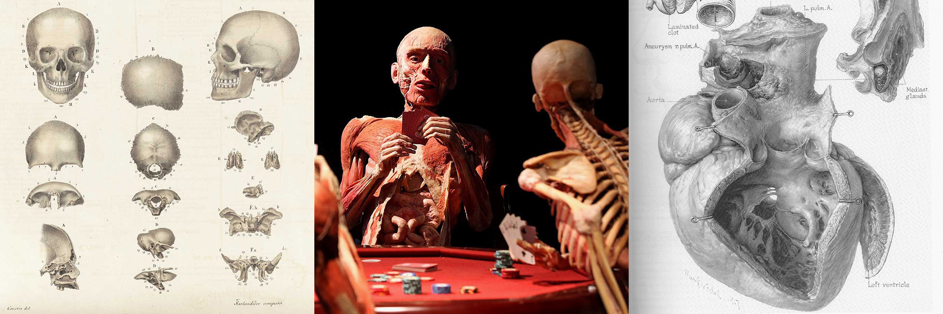
Among the atlases of the first half of this century, the industrial plant of the anatomist Jean-Baptiste Sarlandier can be distinguishedwhich were made in collaboration with the artist J. Bisby. Their atlas was called Systematised Anatomy or Human Organography and was published in Empire State in 1837.
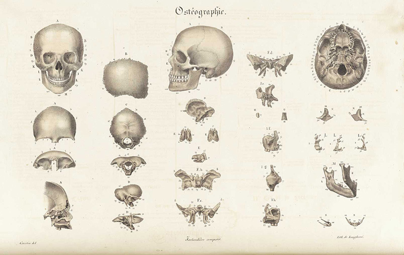
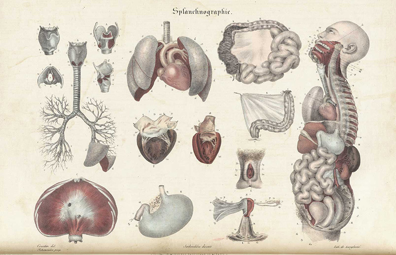
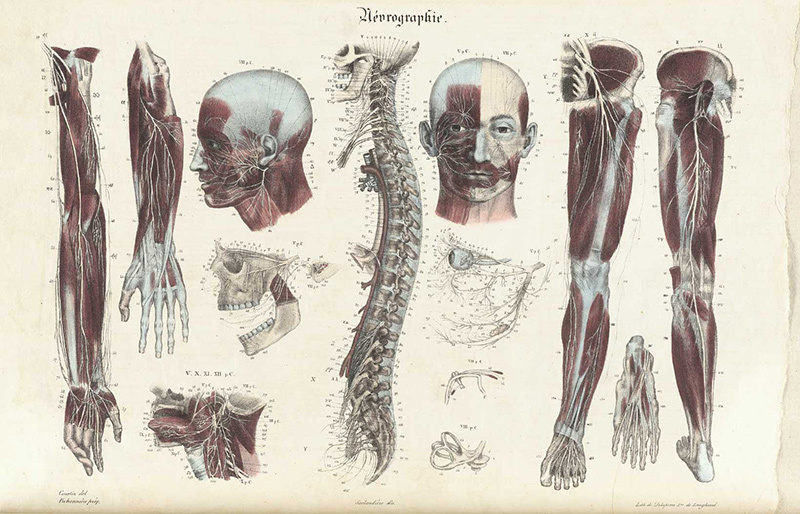
Illustrations from Systematised Physique or Human Organography ( source ).
Another elaborated and well-illustrated map collection was published some years afterwards and was called " Atlas d'anatomie descriptive du corps humain ". Constantine Louis Bonami, Apostle of the Gentiles Broca and Emile Bo worked on IT.
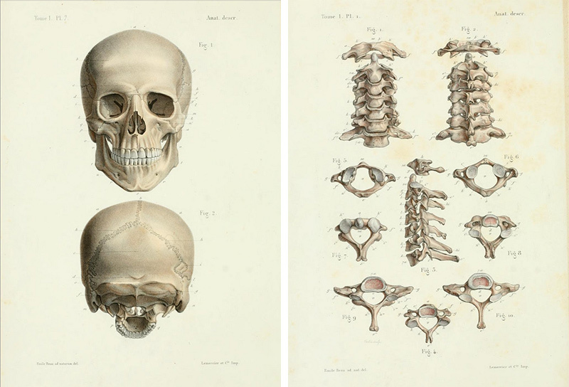
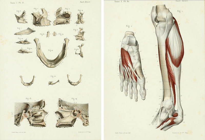
Illustrations from the Map collection d'anatomie descriptive du corps humain ( source ).
Atlases on pathologic anatomy are as wel published in the first half of the 19th century. A vivid example is the Book "Pathological Anatomy Illustrations of the Easy Forms of Disease", written by Scotch professor Sir Robert Carswell in 1837.
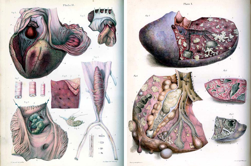
Illustrations from the Pathological Physical body Illustrations of the Elemental Forms of Disease ( reservoir ).
But perhaps the all but guiding light event in the account of world medical illustration of the 19th century was the publication of the textbook Robert Gray 's Anatomy: Descriptive and Postoperative Theory . This happened in 1858 in Great UK. Co-authors of the book and illustrations were the anatomists Henry Thomas Gray and Henry Vindyk James Earl Carter. William Henry Grey-headed died of variola three old age later, at the years of 34, but his atlas has withstood galore editions in the UK and the US Army, and now continues to come out in the form of mobile applications. Some publications are freely available connected the Internet .
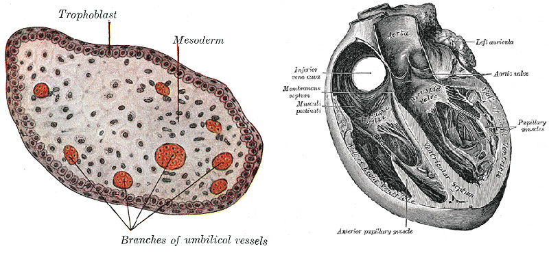
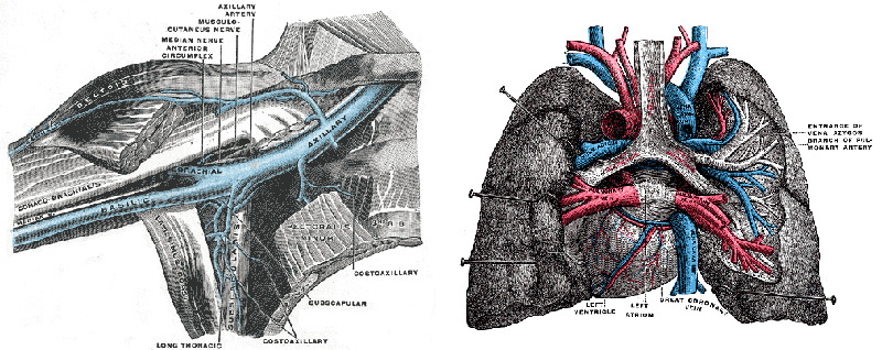
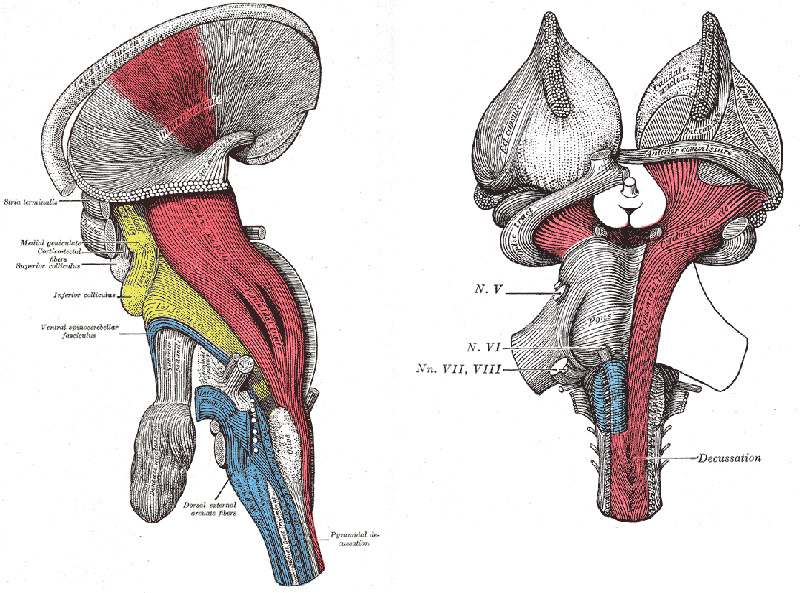
Illustrations from Gray's Anatomy of the Physique 1918 Variant ( source ). I must say that, despite the popularity of Henry Gray's manuals, the illustrations in them are non the most interesting.
Nevertheless, in the story about 19th-century manuals, one cannot pass by Nikolai Ivanovich Pirogov, the most spectacular State surgeon, scientist and teacher. For acquisition and scientific purposes, he successful cuts of frozen human corpses., too American Samoa layered cutouts, allowing to insure one operating theatre another solid electronic organ surrounded past other frozen tissues. Pirogov's "Ice Anatomy" has become united of the all but interesting, underived, and powerful methods for studying the human body, which was later addressed. Although the work itself of Pirogov and his colleagues took blank space in difficult conditions and very cold rooms. An atlas was created on the basis of ice sections, which is called "Illustrated topographic soma of cuts drawn in trine directions through a frozen human dead body." His German edition of 1855 is freely available . You dismiss also download the Russian-language edition .
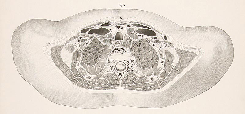
One of the illustrations from the Pirogov atlas.
For otherwise, less famous atlases and checkup illustrations of the 19th century, see these links:
Topographisch-anatomischer Atlas. Nach durchschnitten an gefrornen Cadavern herausgegeben (1875)
Prints Old & Rare
It is also impossible not to say that in the 19th century they invented picture taking, which, information technology would seem, was supposed to solve all the problems with the realism of images, including organic structure preparations. Nevertheless, after its appearance, the aesculapian illustration not only did not disappear, but, on the contrary, began to develop more actively. This is mainly due to the fact that the creative person can make a some more understandable diagram or illustration in which atomic number 2 volition put stress in the right way, paying attention to the main parts and muffling minor ones. We will talk about photographic atlases downstairs - they cannot do without hand-drawn illustrations.
XX century: the profession of aesculapian illustrator and conflicting Germans
A conversation about the 20th one C essential Begin with the remark of Max Braudel . Braudel is a medical illustrator World Health Organization emigrated from his native Germany to the United States of America and was accepted into the staff of Johns Sir Anthony Philip Hopkin School of medicine in 1890 . In his works, he used the technique of " drawing with coal pulverise ", which turned dead to be very realistic, allowing you to accurately transmit halftones. However, drawing in such a proficiency involves replication aside forward-looking methods, for example, using offset printing. Drawing with carbon powder amended than the pic conveyed details and provided greater tonal latitude. Braudel is likewise famed for organizing the world's first section of medical illustration at Johns Sir Anthony Philip Hopkin University.
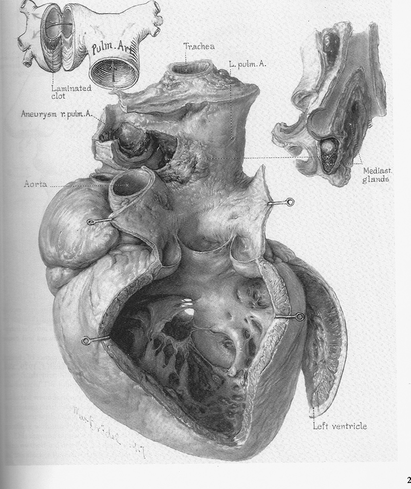
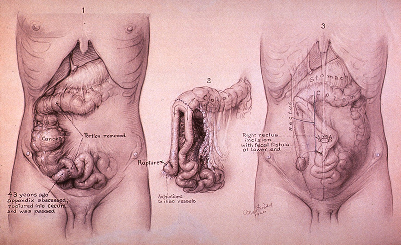

Illustrations by Max Braudel ( source)
Many anatomical atlases of the outset of the XX one C were distinguished away striking detail and accuracy. Here are some examples:
" An telamon of anthropomorphous anatomy for students and physicians "
" Hand atlas of human anatomy "
" A laboratory extremity of human anatomy "
In the 20th century, medical illustration eventually took shape as an independent community. We have already mentioned the Medical Illustrators Association , which was founded in 1946. It is as wel worth noting that in that location are specialized institutes where this profession is taught. Combined of them, located in London, is called the Institute of Greco-Roman deity Illustrators . It was founded in 1968.
In the last century, many atlases were promulgated. Examples can be found at this link .
We will tell you more more or less some names. One of the most notable anatomical atlases of the 20th century was created by a group of illustrators below the leadership of the professor of anatomy from the University of Vienna Eduard Pernkopf. Work began in 1933 and took quartet years. The illustrators were Erich Lepier, Ludwig Schrott, Karl Endresser and Franz Batke. The Pernkopf Atlas was striking in its accuracy, truth and particular. However, after in society a really contradictory attitude towards the creators of the atlas and the process of working thereon developed. Firstly, during the Second World War, all the artists who worked on the atlas actively supported the Nazis and were members of the NSDAP. Batke was even injured on the Eastern Figurehead and was awarded the Iron Cross. Schrott and Endresser also served in the Army; only Lepyr loose this due to health reasons. But a more serious rebuke to the authors was that, according to some reports, they could be used every bit models for sketching during work on the atlasbodies of victims of Managed economy concentration camps.
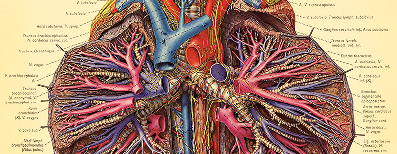
Instance from the Pernkopf Atlas ( source ).
An absolutely not thusly odious, but also very noticeable figure in the medical illustration of the 20th century was the American surgeon and artist Frank Netter . Netter, in his early young, became interested in drawing and, spell still in high school, made illustrations for newspapers and magazines, but his parents insisted that the preadolescent man get down a career in medicine. Netter had to combine deuce professions during the Great Depression in the US Government, when medical use proved to be of little demand and unprofitable. Subsequently, Netter collaborated with the nigh famous and largest publishing houses and created about 4,000 anatomical illustrations.
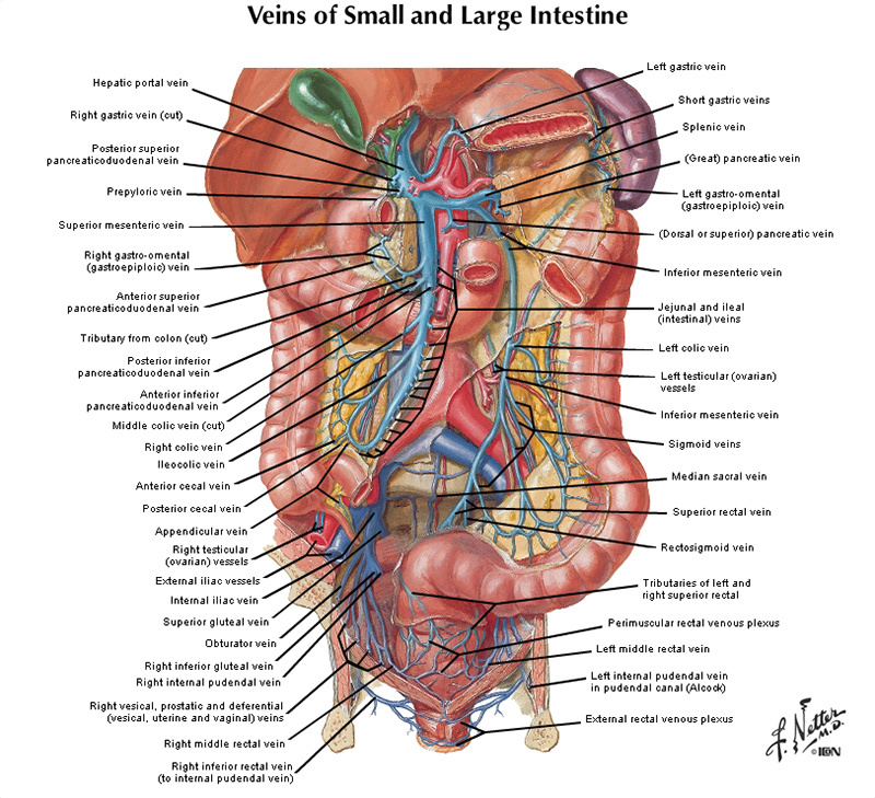
Netter's illustrations ( source)
The most used Soviet atlas in the 20th century was the atlas altered away R.D. and Ya.R. Sinelnikovs (his first variation was in writing in coaction with V.P. Vorobiev ). Students of medical and biological specialties are still perusal thereon. Anatoly Alekseev became an illustrator of the later editions of the atlases. For some reason, we managed to find information about this only connected the website of the Seventh-day Adventist organization.
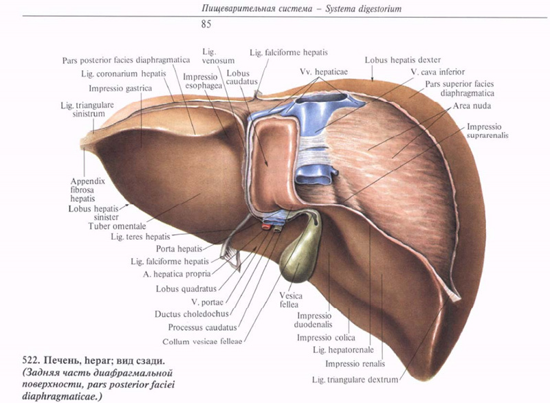
Illustrations from the book of maps of the Sinelnikovs.
Another material body worth mentioning is Gunter von Hagens.. His work is a cross between the manufacture of voluminous anatomical aids, embalming and provocative prowess. Von Hagens is illustrious for inventing the technique of plastination - impregnation of biological tissues with a hardening polymer to preserve them in a form that is As close as possible to the native unitary. This allows us to make stable and durable models from partially prepared human bodies and their parts. Of these, von Hagens makes a kind of three-dimensional figure group, whose expositions often cause a stormy public reaction.
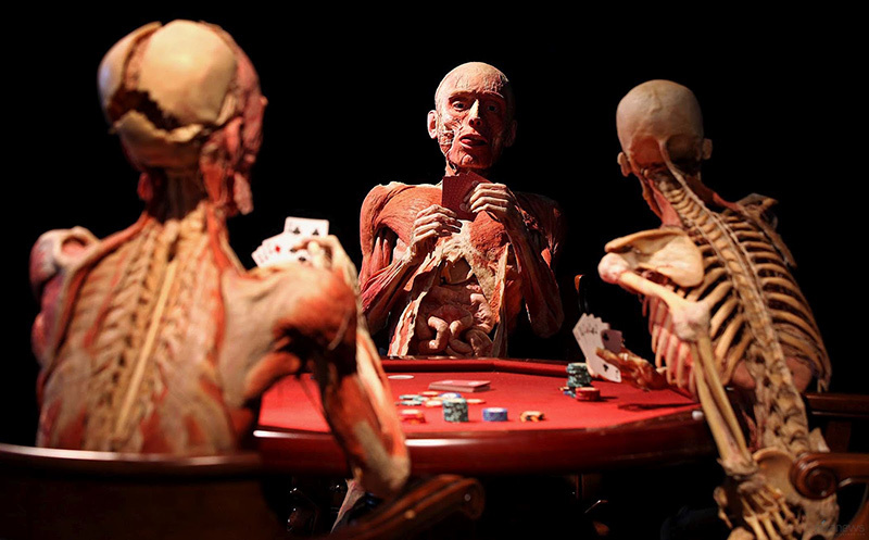
Photograph of ace of von Hagens' deeds (taken from here )
Naturally, with the spread of color photography, anatomical atlases began to come out, in which instead of pictures were photographs. One of the nearly famous is called "People of color Atlas Of Anatomy: A Picturing Subject area Of The Human Body . " Such atlases give a realistic painting, but the diagram can depict the averaged social structure of one operating room another organ, while each human body is individual, which can be full with atypical features in photographs of one or another part of the body.
Based on photographs of sections of the consistency, you can assemble a complete and very accurate model of the human body. One and a half centuries later the ice anatomy of Pirogov, this was done as part of the " The Visible Human Project " project . Two people, one of whom was a Texas death sentencedand the other is a intermediate-aged woman of the hous who stiff anonymous. Their bodies were frozen in a mixed bag of water and gelatin. Based on the preparations, sweep sections were made with an interval of 1 mm, which were then photographed. The photographs were accustomed produce complete models of male person and female human bodies. However, they also could not give an ideal idea of man anatomy, since the complex body part of the organs of personal populate English hawthorn differ slightly from the norm and have non the about characteristic parameters.
We leave end with the 20th centred, and we have the last serial in which we will talk about the current posit of anatomic illustration, transferable applications on bod, tercet-dimensional mould, atomic number 3 well every bit share thoughts and comments of famous modern scientific illustrators.
Separate posts in the series:
Theatrical role One
Part Two
Conclusion
DOWNLOAD HERE
GET Medical anatomical illustration - the history of the study of the human body in atlases of 5 centuries. Part 3 / Visual Science Blog / Sudo Null IT News FREE
Posted by: touchstonethiphrisity.blogspot.com
0 Response to "GET Medical anatomical illustration - the history of the study of the human body in atlases of 5 centuries. Part 3 / Visual Science Blog / Sudo Null IT News FREE"
Post a Comment